 Plateforme de génomique, IPMC UMR7275 660 Route des Lucioles, SOPHIA ANTIPOLIS, 06560 VALBONNE tél: 04-93-95-77-77, fax: 04-93-95-77-08 |
|
Expertise
La plateforme de génomique fonctionnelle de Nice Sophia Antipolis existe depuis 1999.
Initialement orientée vers la conception, la fabrication et l'analyse de puces à ADN, elle a contribué à ouvrir cette nouvelle technologie à
une large communauté, mettant à cette occasion en place un système d'information performant (Mediante),
capable de gérer de grandes masses de données, et fonctionnant en production depuis plus de 20 ans.
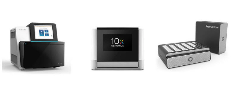
Equipements
Les résultats sont stockés automatiquement sur le portail d'informations de la plateforme Mediante. Cela concerne notamment les fichiers .BAM d'alignement, les fichiers .BW de couverture et l'ensemble des fichiers de l'analyse secondaire et des analyses statistiques conduites en partenariat avec le collaborateur. Sur demande l'ensemble des données brutes sont également mises à disposition et une aide est fournit pour la soumission des données vers la base de données publiques GEO (Gene Expression Omnibus). |
Related publicationsPonzio Gilles |